Explore 2019 Research
Internet-based management for rotator cuff tendinopathy
| Funded by: | Reckitt Benckiser (Nurofen) |
| Recipient: | Professor Peter Malliaras |
| Intended Department: | Department of physiotherapy, Monash University |
| Project: | Internet-based management for rotator cuff tendinopathy |
Plain language summary (Lay report)
The information you provide here will be used to communicate the ‘story’ of this research to a non-scientific readership, so please assume no background understanding of the research area or of scientific concepts in general. Please use plain language, avoiding scientific terminology. If this is unavoidable, please provide plain language explanations. It usually isn’t necessary to explain scientific acronyms or the logic behind the naming of molecules.
We ask that you pay special attention to placing the research in its wider context – how the research contributes to the understanding of disease and why this is important, or how it contributes to the development of a solution to a clinical problem. Although challenging, this is particularly important for basic laboratory research.
a) Give a brief overview of the research you have undertaken, what you hoped to achieve and what you have achieved. Your answer should be no more than two or three paragraphs.
We tested online internet based and telehealth delivery of first line care for a specific type of shoulder pain known as rotator cuff related shoulder pain. This is the most common type of shoulder pain and people are often affected for many months. We know that people with shoulder pain do not always get high quality first line care which includes advice and exercise. Delivering care via the internet and telehealth may help to reach people in need more effectively, so they get the care they need at the right time. This initial study funded by Arthritis Australia was a pilot investigating the feasibility of a trial testing advice only (control group) compared with internet delivered care, and internet delivered care supplemented by telehealth (videoconference sessions with a physiotherapist).
The pilot trial showed that it is feasible to deliver care for shoulder pain via telehealth. People attended the telehealth sessions and undertook the exercise they were prescribed. They also reported that the interventions were acceptable and helpful. The internet only group showed mixed adherence as some people did not undertake the exercise they were prescribed. We have gone in to win two grants for full-scale trials that are designed to test whether our internet and telehealth supported interventions are actually better than control. The first of these trials is due to be reported in 2024.
b) What questions did the grant set out to answer? What problems did you try to solve, or gaps in knowledge did you try to fill? Why is this important? Your answer should be at least 100 words.
The main question was whether our online only and online supplemented by telehealth interventions would be feasible for participants. That is, would participants actually do what was intended and would they find it acceptable. We were also interested in whether there would be any adverse events. Assessing feasibility is very important when testing an intervention that is different to how care is delivered usually. Online only and telehealth is not standard care for people with shoulder pain. It was important to know whether this type of care could work prior to engaging in a large and expensive trial.
c) What did you discover during the course of the grant? Your answer should be at least 200 words.
We had several feasibility outcomes that we tested. This included how fast we could recruit people, how many people stayed in the pilot trial for the full 12 weeks, adherence to exercise interventions, how many adverse events there were and how many of these were serious. We found that we had to screen 68 potentially eligible participants via telehealth to recruit the 36 participants for the trial (12 per trial arm). This took a total of 1 month. At the end of the 12 weeks 34 of the 36 participants completed outcomes, so 94% of people remained in the trial. Adherence to the exercise was very high in the group that had telehealth. 92% of this group did their exercise 2 or more times per week. The internet only group had a lower rate of adherence. Only 67% did their exercise 2 or more times per week. There was only 1 serious adverse event – a participant in the recommended care with telerehabilitation group had elective surgery (brain surgery unrelated to shoulder pain). Other adverse events in all 3 groups could be categorized as non-severe (e.g. muscle soreness, or back pain). Overall, we found that for all the trial feasibility outcomes, all were acceptable aside from adherence in the internet only group.
d) Have the findings of the research already benefitted people with musculoskeletal disease? How might the findings inform further research to help sufferers in the future?
The findings of this research have led directly to a full-scale randomised controlled trial. We have completed recruitment of 346 people for this trial and will report the findings in 2024. This trial will provide a definitive answer regarding the safety and effectiveness of telehealth and internet delivery of care for shoulder pain, which has the potential to provide greater access to care for millions of people in Australia and internationally who suffer shoulder pain.
e) Are you planning to continue the research? Please provide details.
We are excited to have won two major grants provided the finds needed to further develop and test our online and telehealth interventions. The latest of these will test our interventions against usual care. This work will tell us whether online and telehealth interventions can provide benefit for people with shoulder pain.
Scientific summary (Scientific report)
a) What were the main scientific objectives of the grant?
The primary aim of this study was to assess the feasibility of a future substantive randomized controlled trial (RCT) comparing the effectiveness of three internet-delivered interventions for RCRSP (advice only, recommended care, and recommended care with telerehabilitation). Primary feasibility aims included assessing (1) rates of conversion and recruitment, (2) levels of adherence, (3) rate of retention, (4) incidence of adverse events, and (5) response rates to questionnaires.
b) What were the main scientific achievements of the grant? Your answer should be at least 200 words.
Rate if conversion and recruitment
The total cost of the Facebook advertising campaign was Aus $2,146.02 (US $1512.94). The campaign was active for 21 days over 4 weeks—average cost of Aus $59.61 (US $42.03) per participant recruited)—which generated 71,201 impressions and 2492 clicks to the trial site. There were 68 potentially eligible participants who were screened via teleconference, of which 38 were eligible and 36 consented to participate (rate of conversion=36/38=95% of eligible participants recruited). All 36 participants were recruited in 1 calendar month.
Retention and email questionnaire response rates
The questionnaire data at 12 weeks were submitted via email by 34 participants (34/36, 94% retention). Response to email questionnaires at 4 weeks (32/36, 88%), 6 weeks (32/36, 88%), and 8 weeks (30/36, 83%) were also acceptable.
Exercise and telehealth adherence
Acceptable exercise adherence was achieved in the recommended care group (11/12, 92%) but not without telerehabilitation (8/12, 67%) (Figure 3). One-third (4/12, 33%) of people in the recommended care without telerehabilitation group performed no exercise at all. A mean of 10.6 (SD 1.4) out of 12 (88%) teleconference sessions were attended.
c) What problems, if any, did you encounter in achieving the project’s objectives, and how did you address them?
The main issue we encountered was the workload associated with developing the online intervention. This involved developing cartoon videos with voiceover providing education and advice to patients. There were several drafts if the storyboard and substantial editing of the videos required to produce a final product that the research team was satisfied with.
d) Have you disseminated, or plan to disseminate, the results of this research? Please tell us about:
- References for peer-reviewed papers that have been published (please provide pdf copies of papers if possible)
Oral health and rheumatoid arthritis
| Funded by: | HJ & GJ Mackenzie Trust & Arthritis Australia |
| Recipient: | Ky-anh Nguyen |
| Intended Department: | Institute Dental Research- Westmead Hospital |
| Project: | Antibody Cross Reactive Epitope of P. gingivalis |
 For many centuries, there has been a suggestion that oral disease may increase the risk of acquiring (rheumatoid) arthritis – some recent epidemiological evidence supports this association. Why oral disease increases this risk is not well understood and our work seeks to explain the connection between gum disease and rheumatoid arthritis through the involvement of a particular oral bacterium implicated in gum disease.
Rheumatoid arthritis is characterised by the production of antibodies which attack the body’s own protein components. The bacterium Porphyromonas gingivalis is the only oral bacteria known to be capable of producing similar protein molecules to human proteins which are targets of the auto‐ antibody response in many rheumatoid arthritis patients. It has been thought that in people with gum disease and P. gingivalis infection, an inflammatory response in the gums against this bacterium may result in the production of antibodies against the bacterial proteins that also cross‐reacts with similar proteins in human tissues in susceptible individuals, leading to progressive destruction of the joints. The missing piece to this explanation is the unknown protein targets in P. gingivalis that the auto‐antibodies in rheumatoid arthritis are directed against. This project hopes to address this gap by identifying these protein targets in P. gingivalis to confirm this mechanistic link between gum disease and rheumatoid arthritis. The bacterium Porphyromonas gingivalis has been implicated as a leading causative organism for gum disease and these patients often have elevated antibody against the organism.
We have developed a method to quantify the antibody response against the organism and have screened over 300 sera samples from rheumatoid arthritis patients to determined exposure to the bacterium. Currently, we are carrying out statistical analyses to determine whether the level of antibody response to P. gingivalis correlates with various severity markers of rheumatoid arthritis.
This will inform clinicians of the need to reduce exposure to P. gingivalis through the treatment of gum disease as part of the management strategy for rheumatoid arthritis. Using sera with high antibody titre to P. gingivalis, we have developed a technique to detect proteins from the organism that is reactive to the auto‐antibodies in these patients. From preliminary data, auto‐antibodies from each patient binds to approximately 7‐12 distinct bacterial protein targets. Currently, we are screening through the high titre sera to determine which P.gingivalis proteins are most commonly detected between different patients, thus, may represent a common risk marker for a subgroup of rheumatoid arthritis sufferers. Further work will determine the exact identity of these bacterial protein targets to confirm the mechanistic link between gum disease and rheumatoid arthritis. By knowing the bacterial target(s) of the auto‐antibodies in rheumatoid arthritis, it is envisaged that a screening kit will be developed whereby a subgroup of rheumatoid patients be identified, based on their reactivity to the bacterial target(s), to require aggressive treatment for their gum disease to reduce exposure to P. gingivalis.
As auto‐antibodies are detectable in people up to 10 years before the clinical onset of rheumatoid arthritis, the early detection of anti‐P. gingivalis auto‐antibodies may allow for better prevention of RA through improved oral health or allow better control of RA treatment. The preliminary data accumulated in the study will be instrumental in securing larger national grants to continue with the work. We will complete the screen with high‐titre RA sera to compile a list of P. gingivalis proteins that are commonly reactive to auto‐antibodies in RA patients. This knowledge will be vital for the development of any future diagnostic kit to identify at‐risk patients for RA. We would like to thank Arthritis Australia and the generous gift from HJ & GJ Mackenzie Trust to support our work. It is an honour to be given the opportunity to move the research forward and we pledge to ensure the most effective use of the funds given.
3D printing of bio-glue to repair cartilage
| Funded by: | Jointly funded by Eventide Homes Arthritis South AustraliaArthritis Australia – Suzette Gately donation for OA |
| Recipient: | Dr Claudia Di Bella |
| Intended Department: | Department of Orthopaedics-St Vincent’s Hospital – Melbourne |
| Project: | 3D Bioprinting of Bio-adhesive scaffold for cartilage regeneration |
 Musculoskeletal conditions – the suite of injuries and diseases of the bones, muscles, and connective tissue – account for 15.3% of the global burden of death and disability in Australia, second only to cancer (16.2%). For people who injure their articular cartilage, that all important tissue that cushions our joints and enables their smooth movement, a cascade of impacts results from the tears or other physical damage caused by mechanical stresses on the joint. Years of chronic pain, disability, distress, and a reduced ability to work or enjoy physical activity are common implications; trauma to joints causes 14.1% of all serious workplace claims– more than twice that of back injuries. Articular injuries also have severe long term consequences because they almost always lead to osteoarthritis, in itself a major load on the healthcare system, at a cost of $AUD2.2 billion/year, projected to grow exponentially unless something is done to mitigate the condition.
Current surgical options for joint injuries degrade and fail over time, and thus only delay the invasive procedure of a joint replacement, when patients are older and pain is severe enough.
The root problem is that once damage occurs, our bodies cannot regrow normal cartilage. Instead, injured joints normally produce scar like tissue, which neither lasts nor provides the important, movement enabling properties of healthy cartilage. But what if we could trigger the body to instead regenerate the right kind of tissue? To produce not a scar or defect, but normal cartilage, to replace that which was lost? Administered early, such an intervention could transform the outcome of this commonplace injury. Although in the laboratory we can already create tissues that mimic the cartilage present in our body, one of the main difficulties in transferring this to actual surgical implantation is the ability of this tissue to “stick” to native cartilage. Our cartilage is, by nature, very slippery, and nothing can really adhere onto it. Glues can be used, but they damage the cells because they are toxic. The ones that are not toxic are not strong enough to adhere to cartilage.
In this project we have worked to find a special glue that not only allows good adhesion of our printed material to cartilage, but also allows cells to grow in it, so that the new tissue can survive after surgical implantation. We have chosen a glue used in the food industry (with thanks for the idea to Heston Blumenthal!). This material is called transglutaminase.
We have discovered that transglutaminase, when mixed in the right amount and for the right period of time to our gel producing a cartilage (all aspects that we had to understand, define and fine tune in our project), can make our gel sticky enough to the native human cartilage and still let the cells inside the printed gel do their work. These results are extremely important on the path of clinical translation. This approach can now be further optimised to finally be applied in our advanced printing machine that can be used by the surgeon, directly during the surgical procedure, to deliver a gel directly within the cartilage hole in the damaged knee which needs to be repaired. This gel has all the requirements to become cartilage in time. Thanks to our new adhesive properties, this gel will not be washed out after surgery (as it would happen if it was not as sticky), and can remain in place, therefore allowing the cells time to create the new reparative tissue directly in the human body.
I would like to thank the funders of my grant including Eventide Homes, Arthritis South Australia, Arthritis Australia and Suzette Gately’s donation.
Falls prevention for osteoarthritis care
| Funded by: | Arthritis South Australia |
| Recipient: | Prof Ilana Ackerman |
| Intended Department: | Department of Epidemiology and Preventive Medicine-Monash University |
| Project: | Falls prevention: the missing element in osteoarthritis care |
 Osteoarthritis and falls both commonly affect older people. Falls are a leading cause of injury and hospitalisation among older people. In Australia, over 111,000 people aged 65 years or over are hospitalised each year as the result of a fall. The consequences of falls can be devastating for older people and their families, and can significantly impact a person’s ability to live independently. People with hip or knee osteoarthritis are thought to be at greater risk of falling given the impairments associated with these conditions such as pain, reduced muscle strength, and reduced mobility. While we have strong evidence for preventing falls in older people, little attention has been given to managing falls risk and reducing falls in people with osteoarthritis. This is called an ‘evidence to practice gap’.
In designing our project, we hoped to: 1) better understand the risk factors for falls among people with osteoarthritis; 2) examine falls prevention knowledge and practices among physiotherapists who provide care to people with osteoarthritis; and 3) identify factors that make it easier or harder for people with osteoarthritis taking part in falls prevention activities. Our research involved analysing data from a large study of people with osteoarthritis, and we also surveyed 370 physiotherapists around Australia, and conducted interviews with 20 people who have hip or knee osteoarthritis.
This research has generated rich information; importantly it has given us new insights from public health, clinician and consumer perspectives. Drawing all this information together, we have developed a concise checklist for people with osteoarthritis to improve their awareness about falls. We have also developed a checklist for physiotherapists and an information pack to help them assess falls risk in people with osteoarthritis. Building on the research we have undertaken as part of our 2018 Arthritis Australia Project Grant, we are also in the process of developing online training modules for physiotherapists to improve their confidence and skills in providing falls prevention care to people with osteoarthritis.
Balancing muscle force and persistent knee pain in adolescents
| Funded by: | Jointly funded by Arthritis South Australia and Arthritis Western Australia |
| Recipient: | Dr Kylie Tucker |
| Intended Department: | School of Biomedical Sciences, Faculty of Medicine- The University of Queensland |
| Project: | Balancing Muscle force and persistent knee pain in adolescents |
We determined that the balance of muscle force did not differ between adolescents with and without PFP, at any contraction level during either the isometric contractions or during the wall squat tasks. This means that our first hypothesis was not supported. There was no evidence of a difference in the way our adolescents with PFP controlled the force of their thigh muscles compared to the control population.
However, an imbalance of muscle force was observed between the vastus lateralis (VL) and medialis muscle (VM) across all participants during the seated task, which is interesting. In both groups, and at all contraction levels, VL contributed approximately 66% of the total muscle force produced by VL+VM, during the ometric knee extension tasks. In contrast, during the wall-squat VL contributed approximately 53% of total muscle force produced by VL+VM. As such, the force seems to be a much more balanced between the muscles in this wall-squat task. Taken together, these results suggest that the contribution of VL to the total force is different, between the seated and wall squat task.
In conclusion: (i) we provide no evidence that the (im)balance of VL and VM muscle forces differ between adolescents with PFP compared to controls. This may reflect a relatively short duration of disease in our adolescent participants. It remains important to consider the same outcomes in adults with PFP and patellofemoral osteoarthritis. (ii) The wall squat appears to balance the force generation of VM and VL compared to the isometric knee extension task, irrespective of if force is guided through the toes or heels. This has significant clinical implications, as it provides evidence that the way we activate our muscles is altered simply by the type of task being performed.
As this project was partially funded by 2 Arthritis Australia grants, it remains ongoing. The data is in the final stages of analysis, and we will update this section on completion of the additional grant funding. In addition, the participants in our study are now participating in another adolescent patellofemoral pain study, being led by CI-Dr Nat Collins, at UQ. The findings have led to new research questions related to our knee pain population(s). In particular, we do want to continue this work in people with longer duration of pain of muscle activation used during the test tasks. Muscle activation was recorded during (i) seated isometric knee extension tasks (i.e the knee joint angle does not change during these contractions) performed at low, and moderate levels of contraction with the knee at 60º; and (ii) during wall-squats (i.e performing a squat, with the participants back supported by a wall) with force, including body weight, transferred through the toes or heels of our participants.
Knee cap pain in young and middle aged adults
| Funded by: | Funded by Arthritis Queensland |
| Recipient: | Dr Natalie Collins |
| Intended Department | School of Health and Rehabilitation Science, The University of Queensland |
| Project: | Patellofemoral osteoarthritis and predictors of progression in young to middle aged adults with chronic patellofemoral pain |
 Kneecap pain and arthritis are a significant public health problem. Kneecap pain can have a major impact on an individual’s participation in work tasks, caring for children and dependents, and physical activity, each of which contribute more broadly to societal and financial burden.
Kneecap pain affects up to 30% of all adolescents and young adults, and tends to persist and not resolve with time. Importantly, kneecap pain is thought to lead to kneecap arthritis, which is more prevalent, more painful, and occurs earlier in life than general knee arthritis. Young and middle-aged adults with chronic kneecap pain appear to be particularly at risk of kneecap arthritis. Our work has shown that one-quarter of people with kneecap pain aged 26-50 years have signs of arthritis on x-ray, as well as worse function and quality of life compared to those without arthritis.
Because kneecap arthritis has no cure, it’s important that we shift our focus from managing arthritis symptoms to preventing the development and progression of arthritis in at-risk populations. Young and middle-aged adults with chronic kneecap pain may represent an ideal target for arthritis prevention strategies. This research project followed a cohort of people with chronic kneecap pain, who were recruited five years earlier into a study funded by Arthritis Australia. We aimed to determine how kneecap arthritis on x-ray and MRI changes over five years in adults aged 26-50 years with chronic kneecap pain, and how these structural changes relate to changes in symptoms and self-reported disability. Importantly, we also aimed to explore whether factors such as strength, knee alignment and walking patterns – which are potentially modifiable with non-surgical interventions – could predict progression of arthritis on x-ray or MRI over five years.
84 adults with chronic kneecap pain, who were aged 26-50 years, were recruited into the study between 2012 and 2015. They underwent a large battery of tests, including questionnaires, strength and functional tests, alignment measures, x-rays and MRIs of their knee, and detailed assessment of their biomechanics and muscle function during walking, squatting and stair ambulation. At five-year follow-up, 62 (73%) of the original 84 participants completed the online questionnaires, and 45 (54%) underwent x-rays and MRIs of their study knee. The primary reasons that participants did not undergo the x-ray and MRI were a lack of time (e.g. due to work or childcare responsibilities), or because they no longer resided in Australia.
Ethical, referral and funding limitations meant that we were not able to conduct imaging in participants who were located overseas. The x-rays and MRIs are currently being graded by our international radiology colleagues, who will provide expert interpretation of the imaging data. This is the same team who graded the baseline x-rays and MRIs, providing important consistency across the five-year study period.
From the questionnaire data, we see some important findings that demonstrate the ongoing burden of kneecap pain and potential impact of kneecap arthritis. Participants who did not have kneecap arthritis at baseline tended to report the same levels of pain, symptoms, functional impairments and reduced quality of life as they did at baseline (5 years earlier), adding further evidence to the persistent nature of kneecap pain. Participants with kneecap pain who had kneecap arthritis on x-ray at baseline also had persistent pain, symptoms, functional impairments and reduced quality of life five years later, with worsening function on higher level sport and recreational activities. Importantly, the severity of their symptoms was worse than those without kneecap arthritis, both at baseline and at five-year follow-up. This highlights the importance of identifying ways that we can prevent the development and progression of arthritis in young and middle-aged adults with kneecap pain.
Once the x-rays and MRIs have been graded, we will be able to answer the primary questions from this project: (i) how kneecap arthritis changes over five years in adults with chronic kneecap pain; (ii) how changes in kneecap arthritis over five years relate to changes in symptoms and disability; and (iii) whether modifiable factors measured at baseline can predict whether someone with chronic kneecap pain develops kneecap arthritis, or progresses in their kneecap arthritis, over five years. The outcomes of this work will directly inform the development of targeted treatments to stop or slow down the development and/or progression of kneecap arthritis in this younger, at-risk group of people with kneecap pain. We plan to investigate these treatments in larger follow up studies. Because there is no cure for kneecap arthritis, this could be a key step to minimise the prevalence and severity of kneecap arthritis, reduce the personal and societal burden of this common and debilitating condition, and keep young and middle-aged people moving well.
The burden of cancer in systemic sclerosis
| Funded by: | Australian Rheumatology Victoria |
| Recipient: | Dr Kathleen Morrisroe |
| Intended Department: | Department of Rheumatology and medicine- The University of Melbourne- St Vincent’s Hospital |
| Project: | Quantifying the burden of cancer in systemic sclerosis: A data linkage study |
Australia has the highest reported prevalence of systemic sclerosis (SSc) worldwide (233/million cases in 1999). SSc, an autoimmune disease characterised by vasculopathy and fibrosis, is arguably the most devastating of the rheumatological diseases, with a potential to irreparably damage multiple organ systems and shorten life expectancy by an average of two decades. Until recently, the majority of research has focused on the leading causes of SSc-related death, namely pulmonary arterial hypertension and interstitial lung disease. With the emergence of improved treatment and reduced mortality of these manifestations, interest is shifting to the major cause of non SSc-related death, namely cancer.
My research project proposed to quantify the true ‘burden’ of cancer in SSc, including its epidemiology, health service utilisation and impact on health-related quality of life (HRQoL), which had not previously been reported.
Through a data linkage methodology, my project specifically set out to (i) quantify the incidence and prevalence of cancer in SSc; (ii) identify clinical risk factors associated with the development of cancer in SSc, such as immunosuppression and individual organ involvement; (iii) quantify direct healthcare costs associated with cancer in SSc by hospital, emergency department and ambulatory care utilisation and associated cost in SSc patients with cancer; and (iv) quantify health related quality of life in SSc patients.
Our work confirmed that the presence of systemic sclerosis carries an over a two-fold increased risk of developing cancer compared with the background Australian population (standardised incidence ratio (SIR) 2.15 (95% confidence interval (CI) 1.84-2.49), particularly for cancer diagnosed within 5 years of SSc disease onset. Cancer was diagnosed in 14.2% of our cohort, with 43 individuals having more than one cancer type. Specific cancer types with a higher incidence than the background population included breast cancer, lung cancer and melanoma. Additionally, SSc patients with cancer had a higher all-cause mortality than the Australian population, over three fold higher [standardised mortality rate (SMR) 3.13 (95%CI 2.39-4.02)] and higher than SSc patients without cancer [SMR 2.19 (95%CI 1.87-2.54)]. In those with cancer, the primary cause of death was non-SSc related in 58.1%, most commonly due to their cancer (77.1%), followed by ischemic heart disease (5.7%) and sepsis (5.7%). In terms of healthcare utilisation, cancer patients were admitted to hospital more frequently and utilized more ambulatory care services then those without cancer but there was no difference in the frequency of emergency department presentations. SSc patients with cancer compared to those without cancer had an increased healthcare cost with an annual excess healthcare cost per patient of AUD$1,496 (p<0.001). Furthermore, our SSc cohort reported lower health related quality of life than the general population. Cancer patients reported an even lower HRQoL than those without cancer (53.5 vs 38.7, p<0.001).
Our results provide comprehensive cancer data that are essential for designing SSc-specific cancer screening guidelines and for allocation of resources to improve patient care in this devastating disease.
As a clinical researcher I will continue with my research in the field of SSc and hope to share our findings with the wider international community.
Mobile Health applications for monitoring musculoskeletal conditions
| Recipient: | Dr Huai Leng (Jessica) Pisaniello (nee Yong) |
| Intended department: | The Arthritis Research UK Centre for Epidemiology, Centre for Musculoskeletal Research, School of Biological Sciences, Faculty of Biology, Medicine Health- The University of Manchester |
| Project: | The Role of Mobile Health application in real time capture of self-reported symptoms and longitudinal activity, and its feasibility in patient-focused remote monitoring in musculoskeletal disorders. |
Mobile health (mHealth) implementation in clinical care and research has great potential to capture real-time data longitudinally, when compared to traditional epidemiological methods. At present, self-reported symptoms or experience can be captured conveniently and stored digitally within mHealth technologies (e.g. smartphone, smartwatch) and mobile applications (apps). These data, although structurally heterogeneous, are rich sources of health-related information that could reflect a patient’s health journey.
In people with arthritis and other musculoskeletal diseases, tracking symptoms using mobile health apps can greatly improve our understanding of how these diseases affect people and how symptoms can change from day to day. The burden of chronic pain in arthritis is huge. We know that pain remains one of the most important, and yet, a challenging symptom to manage and treat in patients with musculoskeletal diseases. Chronic pain is a dynamic process and can be unpredictable, particularly for those with underlying inflammatory musculoskeletal diseases and concomitant chronic pain condition. Patients are often asked to summarise their overall pain severity since the last clinic visit, which could be weeks to months. This averaged pain severity may not necessarily reflect the overall pain severity over time and in particular, the fluctuation of pain over time. Under- or over-estimation of pain experience can have a huge impact on treatment decision, especially with biologic prescription and analgesic choice. Longitudinal capture of patient’s pain symptoms will help the clinicians in clarifying the definition of disease ‘flare’ and in guiding the trajectory of pain management. From the patients’ perspectives, being able to have better understanding of their pain patterns and levels of pain variability will allow greater sense of control in managing their pain. My research aims to understand how mobile health is implemented in collecting patient- generated health data, as well as how these temporally rich data are analysed. Specifically, my research aims to examine the long-term and short-term day-to-day pain variability over time in a chronic pain study cohort.
As the recipient of this prestigious overseas fellowship grant year 2018, I had the opportunity to work at the Centre for Epidemiology Versus Arthritis in Manchester, UK under the supervision of Professor William Dixon. This 15-month fellowship in Manchester has set off to an exceptionally invaluable foundation for my first year of PhD. I was working with Professor Dixon and the Cloudy team on a large, UK nationwide, prospective mobile health study called Cloudy with a Chance of Pain. This study, which was conducted in January 2016, aimed to examine the association between weather and pain in people living with chronic pain. Although I was not involved in the setup of this project, I came to learn from many in the Cloudy team about the success of this study, in particular the patient and public involvement and recruitment strategies.
The process of preparing and analysing such a large data set has been a huge undertaking, only made possible with expertise input from many researchers with different background (rheumatologists, epidemiologists (national and international), meteorologist, statisticians, mathematician, PhD student with health informatics background and project manager). As a newly trained rheumatologist, doing this fellowship has certainly honed my skills and knowledge in applied epidemiology and statistics in musculoskeletal research.
In Manchester, as a clinical research fellow in the department, I managed to attend various in- house departmental courses, lectures and seminars. These include a 6-month course on epidemiology, genetic epidemiology and statistics in the first half of year, a 3-month course on ‘Statistical Modelling with Stata’ by Dr Mark Lunt, weekly departmental seminars, monthly applied epidemiology sessions, CfE scientific meetings and journal club. I was able to attend the first Digital Epidemiology Summer School course in July 2018, led by Professor Dixon. As a visiting postgraduate student with the University of Manchester, I was able to attend various postgraduate student-related lectures and courses. For my research, I also took the opportunity to learn programming using R for data preparation and analysis and this was made possible with great support and mentorship from Belay Birlie Yimer, a statistician colleague and Anna Beukenhorst, a PhD student with health informatics background. The IT service at the University of Manchester provides programming courses for the staff and students, which I attended to further improve my programming skills in R and Python.
For my research, using the Cloudy with a Chance of Pain dataset, I have the opportunity to examine long-term and short-term day-to-day pain variability in those with musculoskeletal diseases and to examine the driving factors behind the pain variability. First, I descriptively analysed the population-averaged pain severity and other pain symptoms (e.g. pain impact, mood, fatigue, sleep, waking up tired, morning stiffness, physical activity and wellbeing) over one-month period across different rheumatological conditions such as rheumatoid arthritis (RA), osteoarthritis (OA), spondyloarthropathy (SpA) and fibromyalgia (FM). I also performed similar analysis for participants with comorbid FM in RA, OA and SpA. Secondly, I analysed individual-level pain trajectory in terms of daily pain variability over time and change in pain state over time using different modelling approaches. These analyses are currently in progress and the final work will be disseminated in the form of publications and research presentations at rheumatology conferences. I was able to present the preliminary descriptive analyses of my work at the departmental seminar session in June 2019.
In addition, I am currently conducting a systematic literature review on pain trajectory and pain variability in musculoskeletal diseases and I intend to submit this work in the form of publication. All current and future output from this research will largely form my PhD thesis by publication.
I would like to take this opportunity to acknowledge and to thank Australian Rheumatology Association for this fellowship grant funding, and Arthritis Australia for the opportunity to apply for this fellowship within the National Research Program grants. I would also like to thank Professor Catherine Hill, Dr Samuel Whittle and Dr Rachel Black for their encouragement and support during the fellowship application process. This fellowship has been a rewarding and empowering lifetime experience for me as an early researcher, and I can never thank Professor William Dixon enough for his undivided supervision and mentorship, as well as the Cloudy team and colleagues in the department in Manchester.
The role of social media in community perceptions of rheumatoid arthritis drug therapy
| Recipient: | Dr Helen Keen |
| Intended department: | Fiona Stanley Hospital- UWA school of Medicine & Pharmacology –ARA project grant |
| Project: | Community Perceptions of Rheumatoid Arthritis Pharmacotherapy-an analysis of social media platforms |
Rheumatoid arthritis is a common, chronic incurable inflammatory disease. Good therapies have existed for several decades in the form of synthetic disease modifying anti rheumatic drugs (sDMARDS). More recently biologic drugs (bDMARDS) have been developed, but they are associated with risk and enormous cost ($23,000 per patient per year). While bDMARDs have been a great advance for RA patients who have not responded well to sDMARDs, or who experience severe side-effects, bDMARDs are not necessarily better than sDMARDs for most patients. Worldwide trends have seen an increase in the use of bDMARDs, and a reduction in the use of sDMARDS, resulting in spiraling healthcare costs. Patient perceptions almost certainly play a role in this phenomenon.
This project aimed to use software to explore online social media platforms to see if online discussions demonstrate recurring themes relating to drug therapy of RA that may have positive or negative connotations (sentiment analysis). This will provide insight into community perceptions of drug therapy in RA.
We asked a third party company to extract data from social media websites (such as twitter, reddit, facebook) which contained comments regarding the commonly used DMARDS. The company applied algorithms to determine whether the sentiment for each piece of information posted on social media websites were positive, or negative, and the general topic discussed (side effects, cost etc).
We found that in the setting of social media, comments regarding the bDMARDS were generally more positive than comments regarding sDMARDS; the main reason people commented negatively about a drug was because it wasn’t working well.
This finding is important, as online discussions indicate that the RA community are in general more negative about the conventional, cheaper drugs, and generally indicate they don’t work as well as newer drugs. This almost certainly has the potential to impact how effective patients perceive their drugs to be. Clinicians should be aware of this when working with patients to choose drugs to achieve optimal benefit from DMARDs.
This research hasn’t yet benefited people with musculoskeletal disease, but we are planning to publish this work, and undertake further work in the area. In particular, we are looking in more detail at the themes that people with rheumatoid are posting on social media. This is unprompted information from patients (rather than traditional qualitative research which can be biased by predetermined themes) and may uncover issues of importance to people with rheumatoid arthritis. We also wish to explore the language used by people with rheumatoid arthritis in the social media setting.
Identifying factors that reduce the risk of bone fractures
| Recipient: | Dr Feitong Wu |
| Intended department: | Jointly funded by the Vincent Fairfax Family Foundation and the ARA- The University of Tasmania |
| Project: | Early Life Strategies for improving fracture risk factors throughout life |
Older people who sustain a bone fracture have increased risk of becoming disabled, dying early and having lower quality of life. Stronger bone and lower risk of falls are important for prevention fractures before they happen. The overall aim of my project is to find ways to start reducing risk of fractures much earlier in the life span – in children and middle-aged adults, to prevent fractures throughout life. We are doing this using three studies. The first study will use vitamin D research data from around the world to answer the question of whether giving supplements of vitamin D improves bone density in children who are vitamin D deficient (i.e. higher bone density indicates stronger bones). The second study will determine whether bone development in adults is affected by experiences in the womb and as infants (e.g. breastfeeding, smoking during pregnancy, diet during pregnancy and birth weight) and during childhood (e.g. diet, physical activity and fitness, and vitamin D levels). The third study will test whether middle-aged women who had better balance and stronger leg muscle strength had a lower risk of falls five years later.
These unique studies will: (1) answer whether supplements of vitamin D are effective in improving bone density in children with low levels of vitamin D, (2) identify which early life factors affect bone development in adults and (3) identify risk factors for falls in middle-aged adults. We anticipate that these studies will provide critical information on strategies during early to mid-life to help prevent falls and bone fractures throughout life.
Delivering rheumatoid arthritis drugs through oral liposomes
| Recipient: | Dr Meghna Takelar |
| Intended department: | Jointly funded by the BB & A Miller Foundation and the ARA- The University of Queensland |
| Project: | Targeted oral liposomal microcomplexes for tolerizing dendritic cells in rheumatoid arthritis |
Autoimmune diseases such as rheumatoid arthritis (RA) affect 5% Australians and are a major health burden. Current therapies seldom provide drug-free long-lasting remission, with some treatments increasing patient susceptibility to opportunistic infections. Hence alternative therapies, which would rectify the underlying etiology, are essential for prevention and cure. We have explored strategies for co-delivering small doses of self-antigens (self-tissue) along with drugs in liposomes (tiny fat bubbles) to prevent the immune attack on self-tissue and hence treat RA. Currently, this therapy is provided as injections, which are less convenient than oral delivery from a long-term clinical perspective, especially if we wish to progress towards preventive strategies and treatment of children. Further the oral delivery system tends to maximize delivery to the gut immune system, which is ideal for prevention. With the support from Bruce Miller-ARA 2017 Grant, I have been able to develop oral particles, called microcapsules that can withstand the harsh conditions of the stomach and deliver intact liposomes to immune cells in the gut. The gut immune cells can process the drug they contain to suppress levels of disease-causing cells and increase numbers of cells, which would protect the body. In this fellowship, I aimed to extend the capabilities of these liposomes by designing targeted liposomes that will show preferential uptake by the intestinal cells called M-cells. These cells specialize in directing antigens to immune cells in the gut. This novel strategy will improve the delivery of the therapies to the desired sites in the intestine. I had also proposed an assessment of the particles in mouse models to enable progression to clinical trials.
With support from Arthritis Australia, we were able to procure an instrument (January 2018), which would enable reproducible preparation of our oral particles. Similarly, we were able to secure funding (September 2018) to obtain a microfluidizer instrument to prepare liposomes. Coupling the two instruments has now enabled us to prepare the oral particles efficiently with good reproducibility. I have been able to provide evidence that the microcapsules can protect the drug from the harsh conditions of the stomach, deliver it to immune cells in the gut and provide an appropriate immune response. I have also been able to prepare the targeted microcapsules and I have done initial studies to see how they distribute. In parallel, I have optimized the microcapsule parameters to enable assessment in mouse models of arthritis.
Improving outcomes in Rheumatoid Arthritis
| Recipient: | Dr Mihir Dilip Wechalekar |
| Intended department: | Jointly funded by the BB & A Miller Foundation and the ARA- Flinders University |
| Project: | Improving outcomes in Rheumatoid Arthritis |
We gratefully acknowledge the Bruce Miller-ARA Post-Doctoral Fellowship for 2018, which enabled us to significantly advance our research by employing a research associate and establishment of novel techniques of analysis of synovial tissue (e.g. the Vectra microscopy) which would not have been possible without the support of this grant.
The research methodologies we proposed to employ in this project can be separated into 4 broad techniques: flow cytometry (perfect for assessing new cellular subsets), microscopy (ideal for assessing tissue phenotype), transcriptomic analysis (to assess the complete set of RNA transcripts that are produced by genes of inflammatory cells) and serum cytokine analysis (immune system messenger and inflammatory molecules), which together can be used for the identification of new biomarkers that predict effective responses in different patients.
The 2018 Bruce Miller-ARA call for funding was submitted as a 2-year project, where our first step was to perform optimisation and analysis of flow cytometry to identify lymphocyte infiltrates in the ST and to perform routine Immunohistochemistry (IHC) for these early RA ST samples (see below). As the project progresses into the second year, upon which we will have recruited both pre- and post-treatment samples from the joints of RA patients (as well as matching blood samples from these patients) we plan to correlate clinical disease activity, with the presence or absence of the types of cells identified via flow cytometry and IHC, but also with the serum cytokine analysis (ELISA) and transcriptomic analysis (real-time PCR) biomarkers that might confer response or relapse to treatment.
In 2018, we have made significant progress towards achieving the aims of this grant. These achievement progress milestones include: (1) establishment and implementation of multi-colour flow cytometry to investigate the cellular infiltrates in the tissue from early RA joints; during 2018, we expanded our panel to include novel T cell subsets; (2) we developed- for the first time in synovial tissue- a multi-colour microscopy method with automated computational assessment of tissue phenotype. This is a major milestone for several reasons including (1) RA ST is a finite resource and extracting the most amount of data from each sample is important; (2) Preservation of structural information and identifying cells that express more than one marker is difficult with traditional IHC, but possible with multi-colour microscopy, and; (3) The automation of sample analysis will remove human analysis bias and reliability issues.
Additionally, we have recently used transcriptomic profiling by RNA seq on a subset of the early RA patients to identify unique immune cell signatures that are associated with disease and response to treatment. In particular we have identified alterations in gene expression of gene involved in both T cell and plasma cell.
Importantly, as per the aims of this project, we are now in the process of extending this transcriptional profiling to the remaining patients treated with standard therapies, but also those patients in the study that have been treated with biologic therapies.
Finally, by the end of 2018, we mostly established a multi-colour microscopy technique for the histopathological assessment of ST. We now have the ability to use computational analysis to count the number of different cell subsets in the ST, and to distinguish between samples with different phenotypes. This is a step change in the way that histopathological analysis is performed, and development of this technique will translate into high impact data for this project as well as all future projects in all rheumatology laboratories.
Obesity and psoriatic Arthritis
| Recipient: | Dr Premarani Sinnathurai |
| Intended department: | Australian Rheumatology Association- Project Grant- Rheumatology Department- Royal North Shore Hospital |
| Project: | Adipokines: a link between obesity & psoriatic Arthritis |
Psoriatic arthritis is a chronic inflammatory arthritis associated with the skin condition called psoriasis. People with psoriatic arthritis can experience a wide variety of disease symptoms – some have many joints affected, whereas others have only a few. Some people with this condition experience inflammation at the places where tendons connect to bone, known as thesitis. It is not clearly understood why this is the case. There may be some genetic factors involved, and environmental factors may also play a role. Psoriatic arthritis is also associated with other medical conditions, especially cardiovascular disease and obesity. People who are obese are at increased risk of developing psoriatic arthritis, and have a poorer response to treatment for their arthritis. One possible reason for this is that fat cells, known as adipocytes, produce substances called adipokines which may promote inflammation. We did this study to see whether the blood levels of these adipokines were associated with the pattern of joints affected in people with psoriatic arthritis.
We measured the levels of adipokines in the blood from 39 people with psoriatic arthritis from two hospital clinics. We compared these levels with the pattern of arthritis and found that an adipokine called chemerin was higher in people who had many joints affected by psoriatic arthritis, compared with those who had few joints affected. Another adipokine called adiponectin was lower in participants with many joints affected. To our knowledge, this is the first study which has looked at whether these adipokines are associated with the pattern of joint involvement in people with psoriatic arthritis.
Communicating low back pain research to the consumer
| Funded by: | Arthritis South Australia |
| Recipient: | Dr Jenny Setchell |
| Intended Department: | School of Health and Rehabilitation Science- The University of Queensland |
| Project: | Translating low back pain research: identifying potential hidden harms in health messaging |
 The overarching aim of the research was to contribute to improved management of low back pain, the world’s leading cause of disability. People increasingly use the internet to obtain health information. However, most current online information sources do not meet the needs of individuals with back pain, and are often untrustworthy. Accurate and trustworthy education about this condition has the potential to substantially reduce its excessive burden. To address this issue, we had previously developed a (separately funded) online resource. This was done in collaboration with consumer organisation Arthritis Australia; individuals with low back pain; and leading national/international low back pain experts. Our world-first website involved extensive work to identify high quality information and distilled it into an easily understood evidence-based resource in multiple formats (e.g. consumer and clinician videos, information sheets, quiz) and used evidence-based algorithms to create tailored consumer advice. The website’s key messages were:
- enhancing consumer confidence in managing their condition and making evidence based treatment choices;
- de-medicalising back pain and reinforcing that back pain is a natural part of life for many and can often be managed with early return to activity; and
- engaging consumers in behaviours and attitudes to reduce the burden of pain.
The project funded by this grant used techniques from social sciences to evaluate these key messages; examining the principles, understandings and assumptions that these messages take for granted. It considered how these messages (and their underlying assumptions) might be taken up by consumers accessing the website, and considered any potential negative effects on them. The study complemented a separately funded randomised controlled trial of the website.
Project aims were:
- To investigate the understandings and assumptions that were taken for granted in the key messaging of the website.
- To determine how key messages (and their underlying assumptions) were taken up by participants and to consider any potential negative effects on participants.
- To suggest future directions for the website.
- To contribute to critical broader discussions about the nature of pain and arthritis health messaging.
These aims are highly important for reasons both specific to the website, and more broadly for the management of low back pain.
We used two methodologies to achieve the project aims. We encountered no problems with the implementation of these two study methods and are now considering these two methodologies as separate studies as both produced valuable and different results:
Study 1. An analysis of the key assumptions of the website content (words, images, videos etc) drawing on key social theories. We collaborated with a group of people with different backgrounds to do this: sociology, public health, psychology, occupational therapy, physiotherapy and an ‘expert consumer’ (person with low back pain with social science knowledge)
Study 2. A participatory study where the assumptions were further investigated with individuals with low back pain. This study had two main methods of data collection: 1) we observed and interviewed participants as they first interacted with the website, and then 2) over the next month participants took photographs whenever they thought about the website – these photographs were then shared with the researchers and discussed in a second interview to consider how the website affected participant’s lives.
Predicting knee loading using wearable sensors
| Funded by: | Arthritis Western Australia & Arthritis Queensland GIA |
| Recipient: | Dr Amity Campbell and Tara Binnie |
| Intended Department | School of Physiotherapy and Exercise Science- Curtin University |
| Project: | Predicting knee loading using wearable sensors |
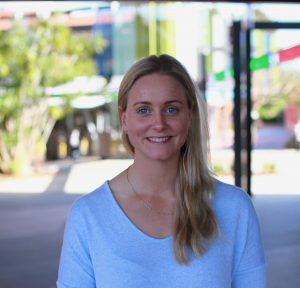
This study aims to validate a model for estimating knee joint loading from wearable sensor data. Currently clinicians and researchers are restricted to using expensive laboratory-based equipment to measure knee joint loading, and this method fails to provide any information on how the patient actually uses their knee in their normal activities of daily living when away from the laboratory/clinic. Several specific measures of knee loading, such as the load acting to push the knee outwards during walking, have been demonstrated to predict the progression of knee osteoarthritis and have been identified as critical measures. Thus, using wearable sensors would greatly advance the understanding of how people with knee OA load their knee in daily life, and this knowledge could assist in the development of effective treatments that slow or stop the progression of knee OA.
Once this research has been completed and it has been confirmed that wearable sensors can accurately predict knee joint loading, we will use the sensors in a range of clinically focussed studies to track how people move in their daily lives. It is hoped that this body of knowledge will better our understanding of the way people with a range of musculoskeletal disorders move and how this associates with their pain and disease progression. The results of this study provides us with great confidence that we will be successful in developing a model to accurately predict knee joint loading in individuals with knee OA, using wearable sensors.
CCL 17 in osteoarthritis and its associated pain
| Funded by: | Jointly funded by Arthritis Australia and Arthritis Western Australia |
| Recipient: | Dr Ming- Chin Lee |
| Intended Department: | Department of Medicine (RMH) University Of Melbourne |
| Project: | CCL 17 in osteoarthritis and its associated pain |
My ongoing research program is to focus on two main goals (i) understanding the role(s) of inflammatory cytokines/chemokines in musculoskeletal (MSK) conditions, including inflammatory arthritis and osteoarthritis (OA) and (ii) defining and validating new therapeutic targets for these diseases.
My early study discovered a new pathway where the chemokine CCL17 is an effector molecule of GM-CSF (granulocyte macrophage-colony stimulating factor) and induces via a transcriptional factor, interferon regulatory factor 4 (IRF4), which is essential for inflammatory and arthritic pain and disease development in experimental animal models. I have also validated CCL17 as a potential therapeutic target using a therapeutic antibody-based protocol. This body of work has been published in a top ranking clinical journal, Journal of Clinical Investigation. I also found that TNF (tumour necrosis factor) is only required at the early stage of disease development (published in Journal of Clinical Investigation Insight). This finding acts as a knowledge foundation warranting future research in other MSK conditions, where pain is relevant. As such, using an experimental model of OA, I have determined the importance of CCL17 in OA pain development and validated it as a potential therapeutic target by using a monoclonal antibody-based therapeutic protocol neutralizing CCL17 in the OA model. This is the first time CCL17 has been proposed as a potential therapeutic target for OA and was recently published in Arthritis Research & Therapy.
Walking for knee osteoarthritis
| Funded by: | Arthritis Tasmania and Eventide Homes |
| Recipient: | Dr Dawn Aitken |
| Intended Department: | Menzies Institute for Medical Research, University of Tasmania |
| Project: | A randomised controlled trial evaluating community walking for knee osteoarthritis |
 Walking is a treatment exercise which is used in the management of knee osteoarthritis, yet there is a lack of evidence about the effect that walking has on the knee joint. Therefore this ongoing study aims to investigate the impact of a 6 month aerobic walking exercise program on knee joint structure of patients with knee osteoarthritis. Knee joint structure will be assessed using MRI (magnetic resonance imaging). We will also examine the effect of walking on participants pain and function levels.
In this study one half of participants are participating in a 6 month aerobic walking program and the other half are receiving usual care only (i.e they will be in the control group). The participants in the walking group walk 2 days/week in a supervised group session and 1 day/week unsupervised at home. Group sessions are tailored to individual fitness levels and led by a physiotherapist or exercise physiologist with experience in prescribing exercise for OA patients.
The pilot study is ongoing. We have telephone screened 172 participants, and 49 of these have attended a screening clinic visit. Of the 49 screened, 40 have been enrolled in the study and randomised. Recruitment has now closed and the final participant will complete the study in December 2019. Three participants have completed the 6-month follow-up, with the other 37 at various stages of follow-up.
We hypothesise that this study will show that weight-bearing exercise, such as walking, is safe and does no harm to the knee joint. Such knowledge would have major implications, as it would dispel the fears around weight-bearing exercise, providing clinicians with the evidence base to confidently prescribe walking and reassuring patients to follow this advice. I would like to thank the grant funders Arthritis Tasmania and Eventide Homes.
Sign up to Arthritis Insights
Regular updates, news and research findings delivered to your inbox:
